


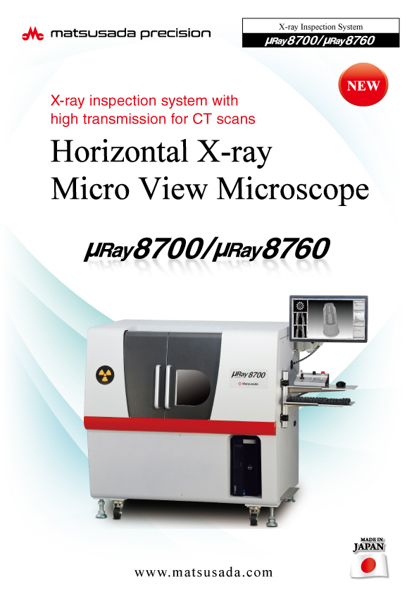
The µRay8700/µRay8760 is a versatile micro-CT system designed to inspect a wide range of objects, from small components to large assemblies. Equipped with a high-power 130 kV, 40 W microfocus X-ray source, the system delivers high-definition 2D images with low noise and 3D reconstructions with exceptional spatial resolution.
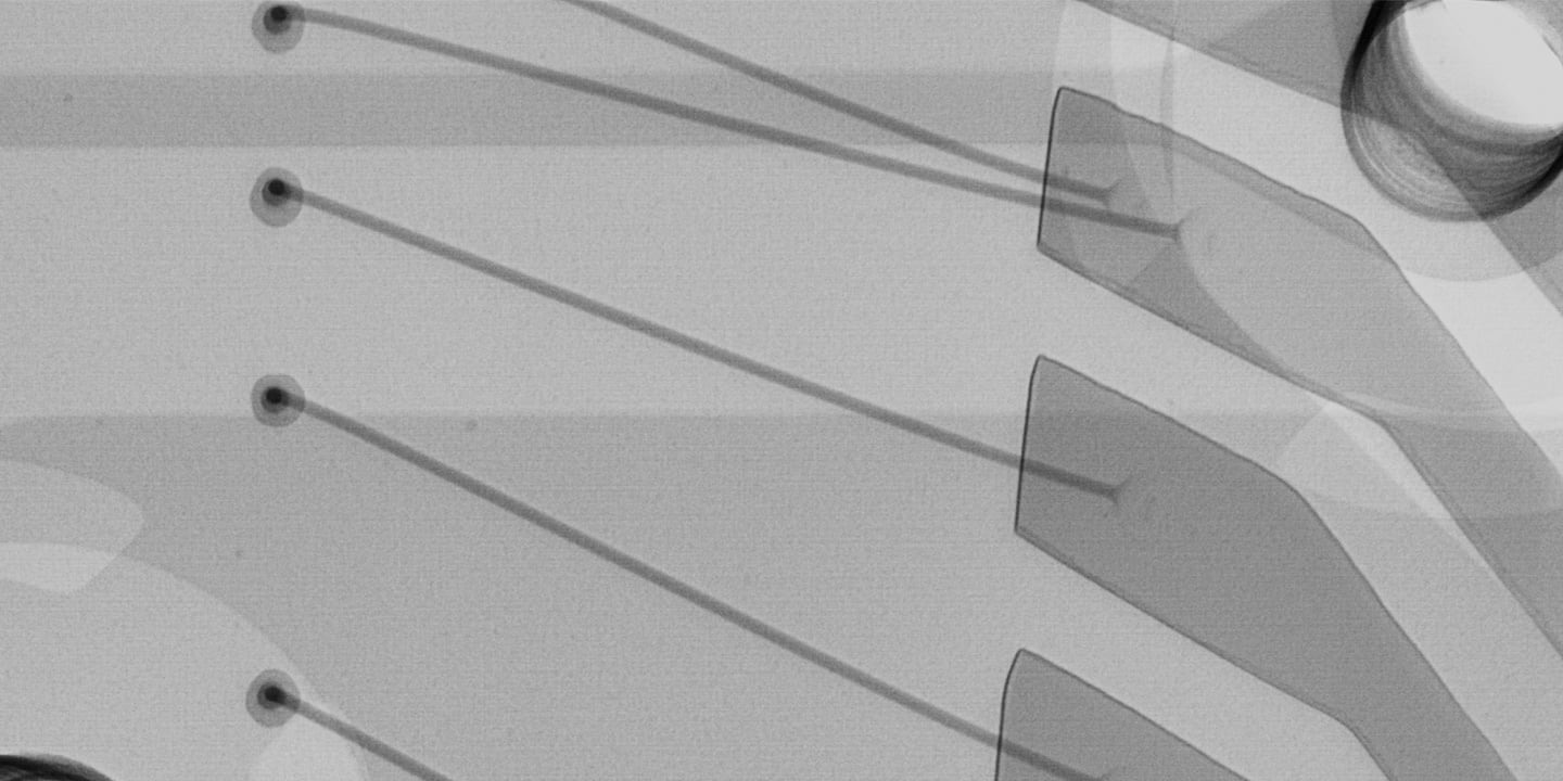
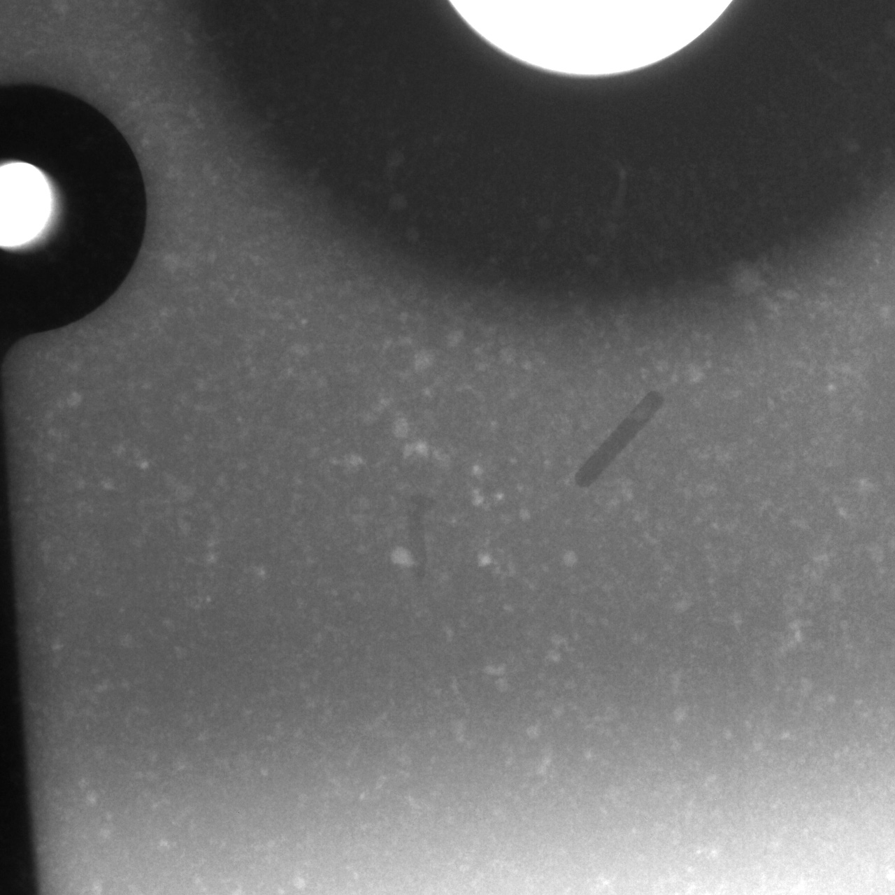
High performance 130kV X-ray tube and high-resolution X-ray camera provide high transmission for clear images.
The exceptional penetration power of the 130kV X-ray tube, combined with high output up to 40W, enables detailed detection of even microscopic defects. The 8700/8760 series offers innovative observation with high transmission.
The overwhelmingly high transmission with 130 kV X-ray tube voltage and the high power up to 40 W provide higher-grade observation with high transmission. We developed our exclusive noise reduction technology as well as the smallest focal spot size of 5 μm with 4 W output. Especially, incredible low noise and high contrast are achieved by the micro-focus X-ray tube. In addition to such superior images, the exclusive software having many effective functions along with superior operability makes μRay8700/8760 series exceptionally high quality system in non-destructive inspection.
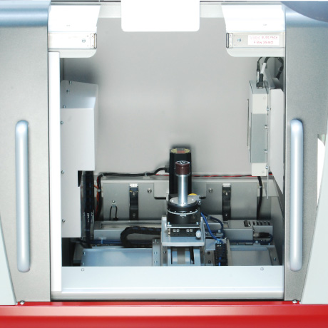
Easily inspect longitudinal cross-sections using the horizontal X-ray irradiation system.
Benefits of horizonatal unit in the large-size stage observation of the entire object.
In addition to the X-axis (left/right), Y-axis (front/rear), and Z-axis (up/down), it also has a rotation axis (turn), which enables
Cylindrical samples can be seamlessly inspected by placing them on the rotary stage for full 360-degree observation.
The stage can be moved to the left/right, front/back, and up/down, allowing for easy operation.
Using a large space stage 3.94 inches (100 mm) in diameter together with the sample fixing platform, the series offers different height settings at work as you need.
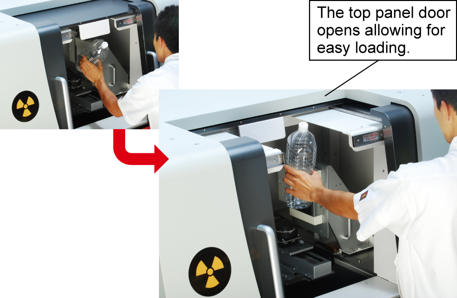
Top Access for Accommodating Long and Heavy Samples
The top panel opens to facilitate the loading of long samples or heavy workpieces up to 10 kg.
* Top panel access is available on the µRay8700 model only.
* Support for 10 kg samples is an optional configuration.
Suited for radiography of powder, liquid, etc. on the rotation stage
The horizontal configuration is ideal for samples that are difficult to inspect using conventional vertical radiography or tomography.
The images will be unclear if a sample position does not stay still in tomography. This system enables radiography and tomography with the sample in an upright position. This minimizes gravity-induced deformation, ensuring stable rotation ideal for high-precision tomography.
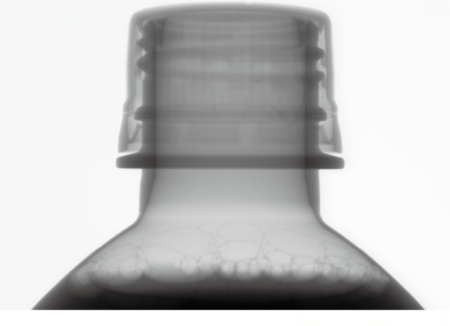
X-ray tube voltage: 90 kV
X-ray tube current: 150 μA
Monitor image magnification: 6.7 x
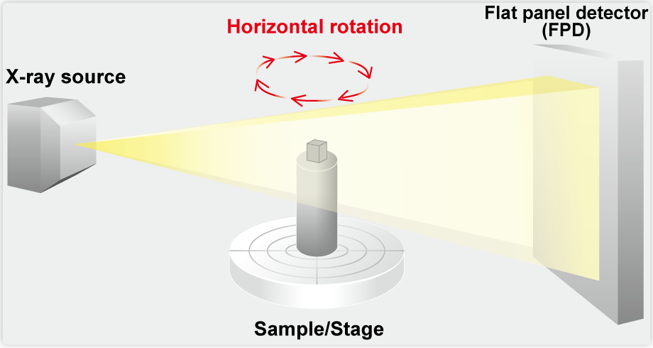
Benefits of horizontal radiation
The horizontal radiation system introduced radiography/tomography with the sample upright. It can ignore the effect of gravity, and the rotation axis is stable during the sample rotating operation, which is ideal for tomography.
Intuitive Operation: Instant access to essential functions [Dedicated Software (Included)]
Allows for simple stage and X-ray control, while offering a comprehensive suite of image processing and measurement functions.
New development
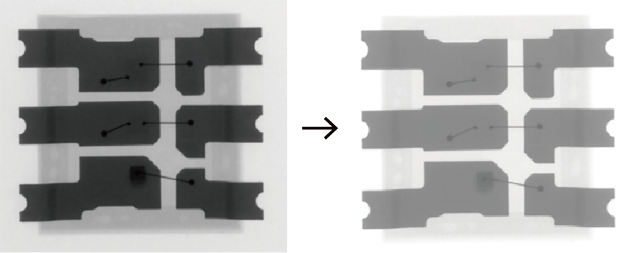
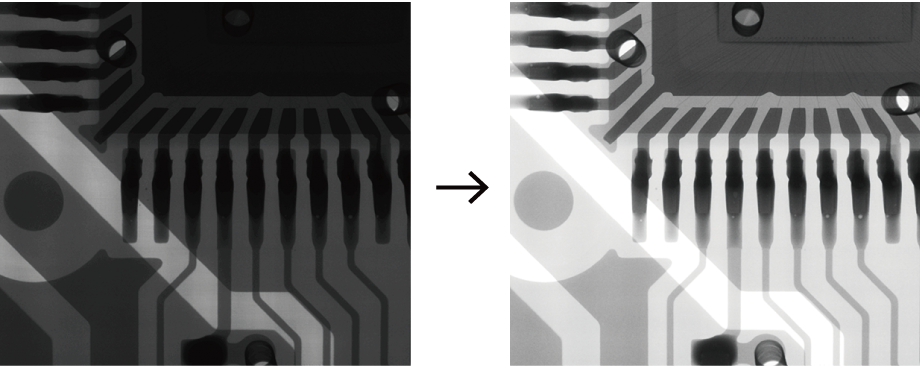
New X-ray image processing function s-HDR
The s-HDR function produces a wide dynamic range of radiographic images. This function visualizes areas that would typically be overexposed (haletation) or underexposed (blocked-up shadows), ensuring optimal visibility across the entire image. You can observe with X-ray images of optimal quality.


Brightness control
Adjusts the brightness of the radiographic images by changing the gain value and tone curve.
Contrast control
The contrast of the X-ray image can be adjusted by adjusting the gamma curve and luminance range.
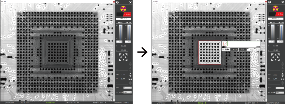
ROI (Region of Interest) function
The area to be viewed can be checked by automatically adjusting the brightness to the appropriate level. For example, it is possible to highlight certain areas of an X-ray image that are difficult to distinguish. Subtle manual contrast adjustment is also possible.
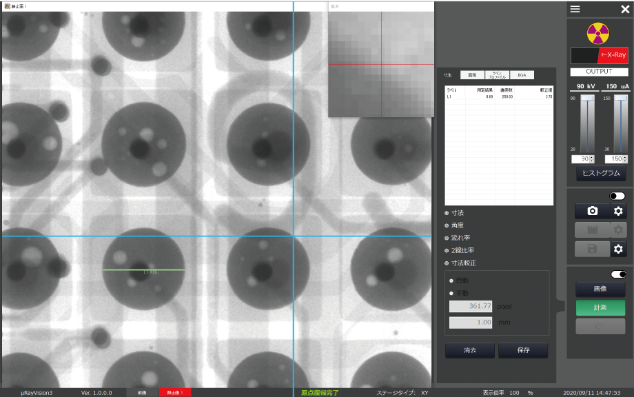
Length measurement
By drawing measurement lines on the captured image, dimensions in the captured image can be measured.
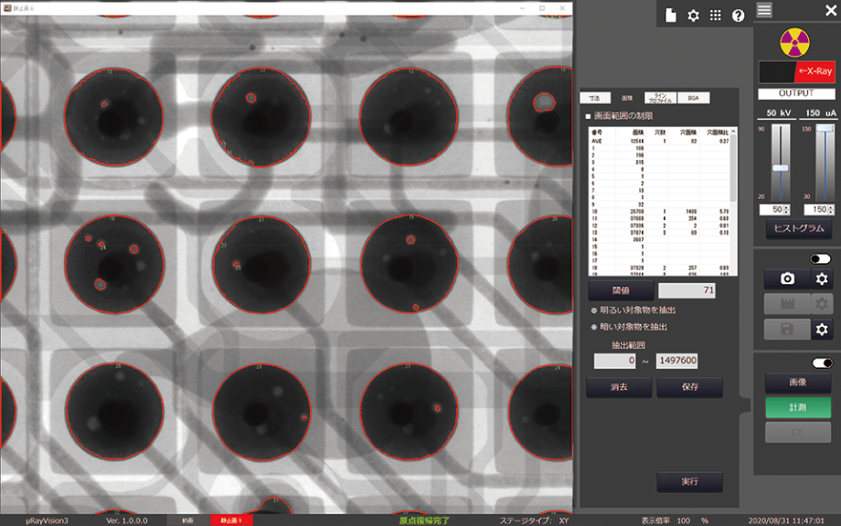
Area measurement
The area can be measured based on gradation boundaries. The measurement result makes it easy to compare samples of a certain shape. For samples such as BGA, it is possible to measure differences in size, void count, void area, and area ratio.
Pseudo color
This function is designed to make it easier to identify specific areas. X-ray images are obtained in black and white tones, but they can be colored. For example, coloring only the components on the PCB makes them easier to identify.
Brightness-specified color
The precision μx8600 has a function that makes it easier to identify defective parts. This function allows the user to specify a range of 16384 shades of gray for defects to be highlighted in color. This allows you to easily see faults that were invisible before.
Black and White image (Threshold/Binary image)
The precision μx8600 has a function that makes it easier to identify defective parts. By setting a threshold, the images are displayed in two colors, black and white.
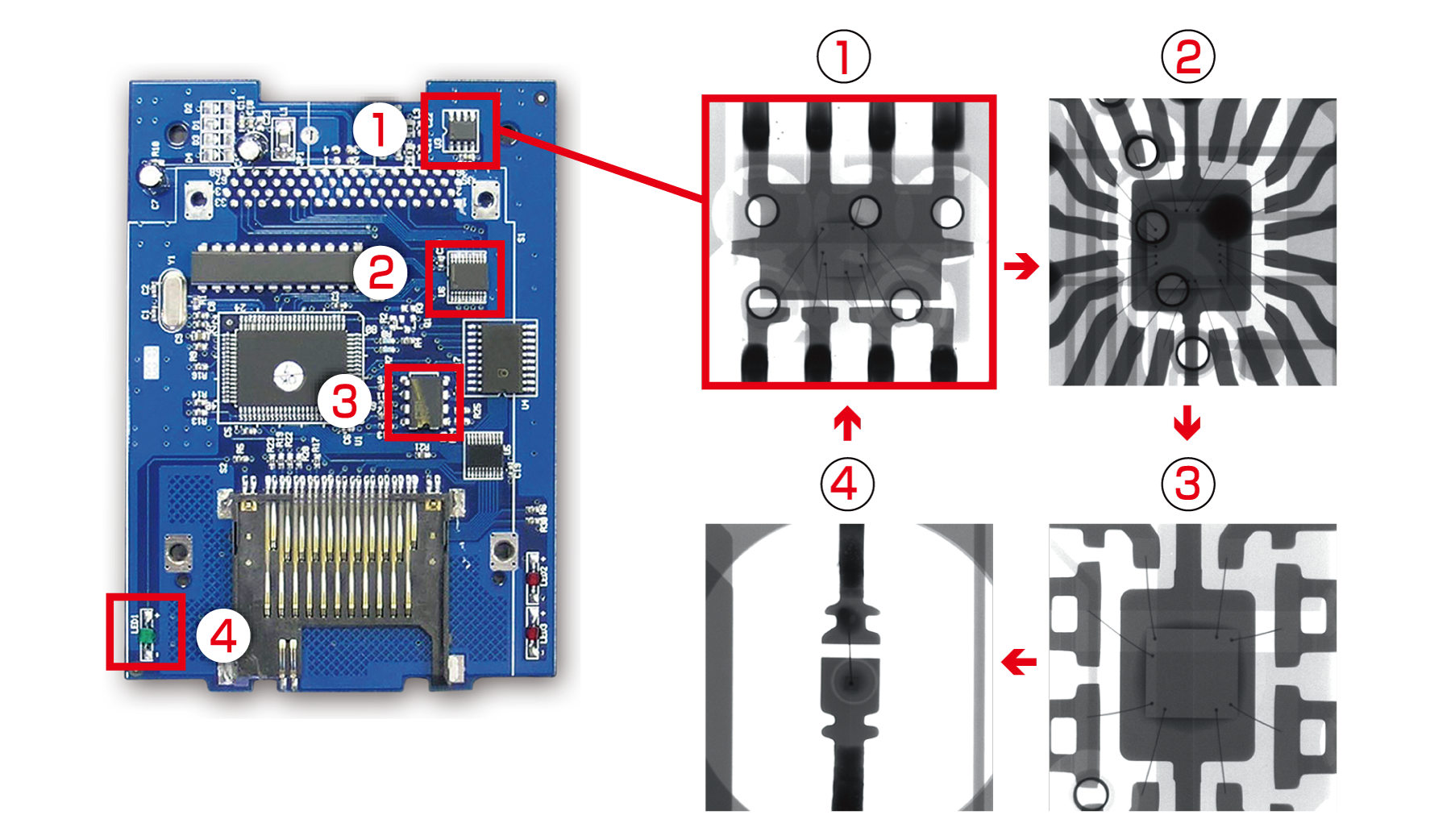
Position memory function
The Position Memory function automatically moves the stage to pre-set coordinates for inspection. Conventionally, the operator had to move the stage and take pictures, but this function makes it possible to take pictures of multiple designated areas with a single click. In addition, the X-ray output value and magnification rate can be set individually for each shooting point, allowing all inspection objects.
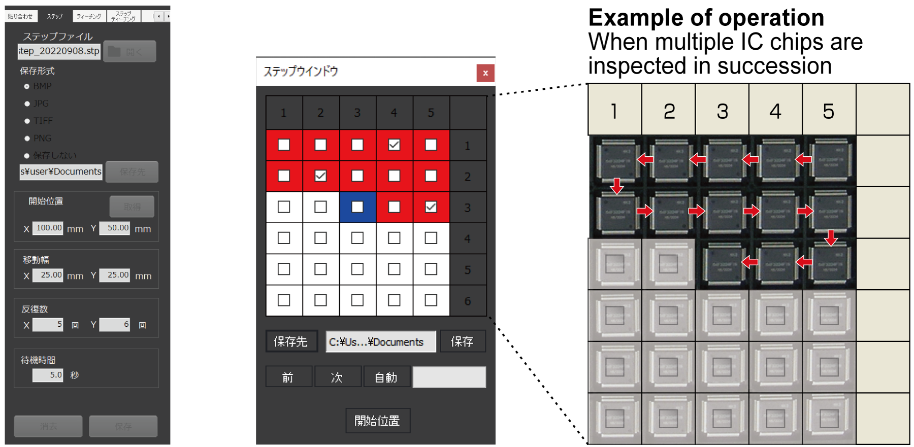
Multiple testing (Automatic feeding)
The continuous inspection can transfer the stage sequentially at a desired pitch to take images. All samples on the pallet can be inspected simply by setting the start position, the movement pitch for each of the X and Y axes, and the number of moves.
High Penetration Power: 130kV Tube Voltage
Capable of inspecting dense metals and thick workpieces.
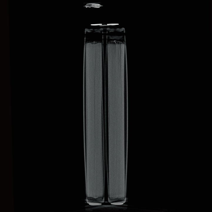
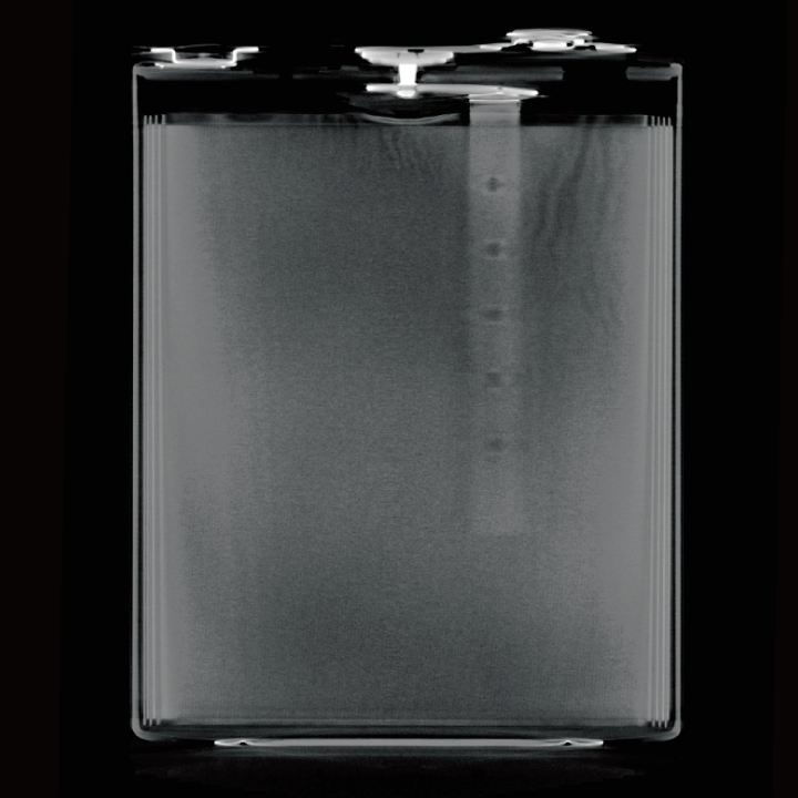
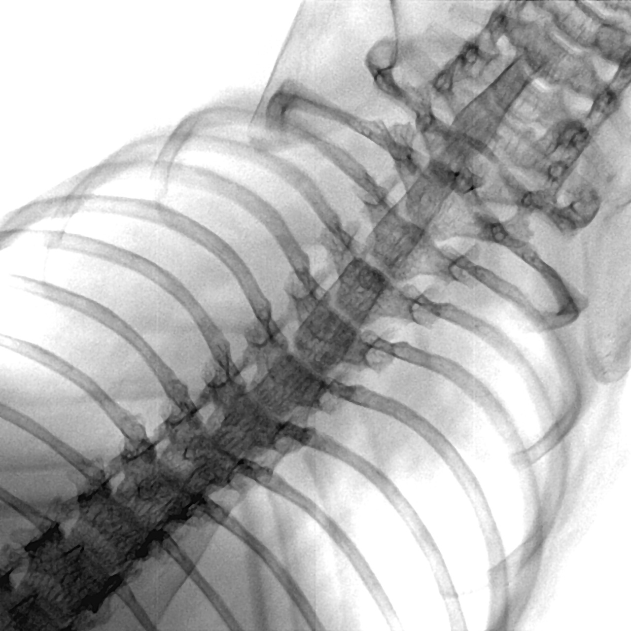
Prismatic Lithium-ion Battery Inspection
Prismatic lithium-ion batteries used in mobile devices, power tools, and small-scale mobility require exceptional safety standards due to their high energy density. Industrial CT systems provide comprehensive quality assurance for everything from individual cells to modules equipped with Battery Management Systems (BMS).
Cell Inspection (Stacking and Welding Quality):Our systems accurately measure internal electrode sheet misalignment and "overhang" (the critical margin between plates) to prevent short circuits. We also detect insufficient penetration in collector tab welds, blowholes, and metallic contaminants to mitigate fire risks before they occur.
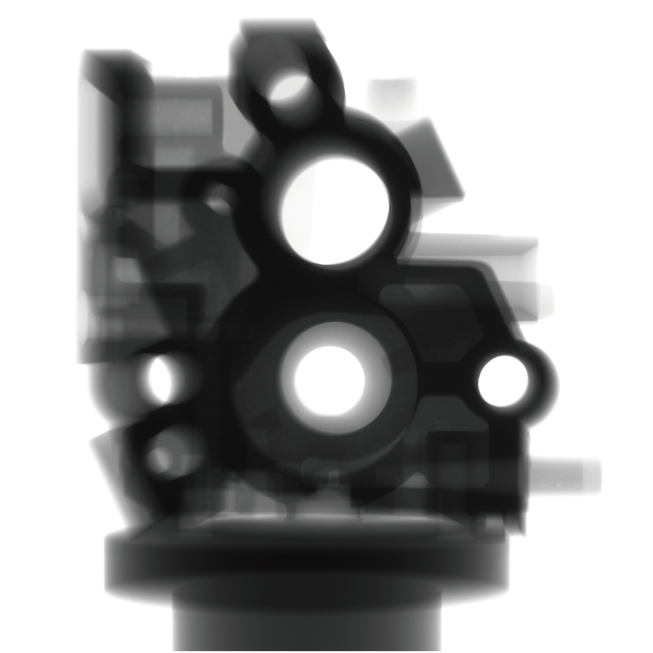
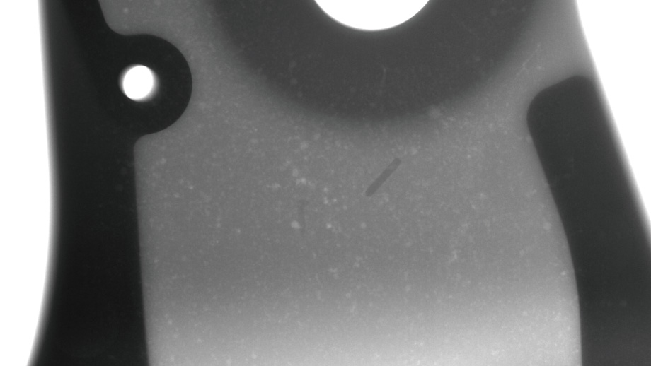
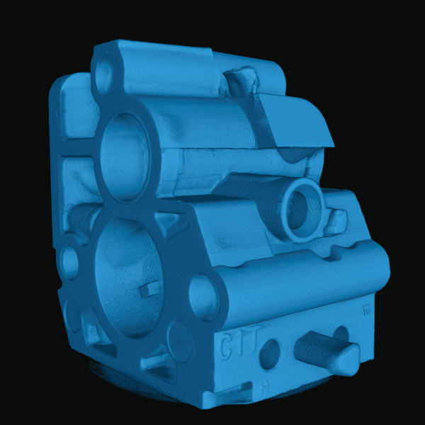
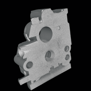
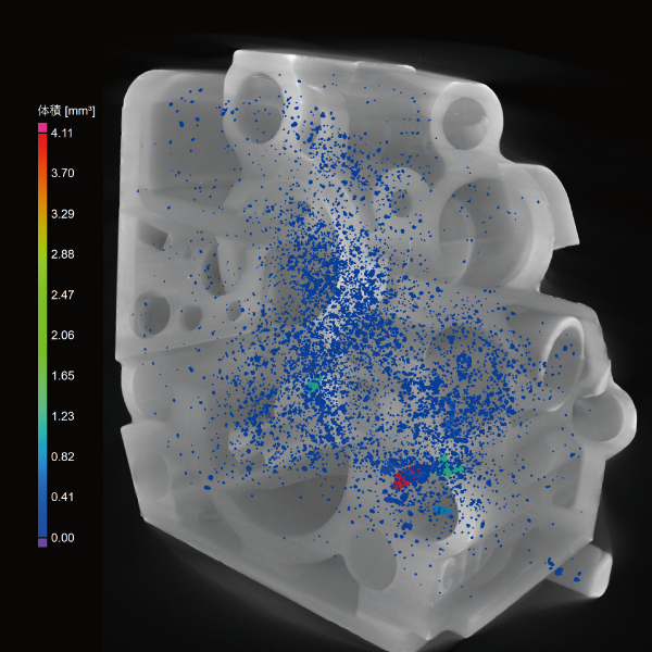
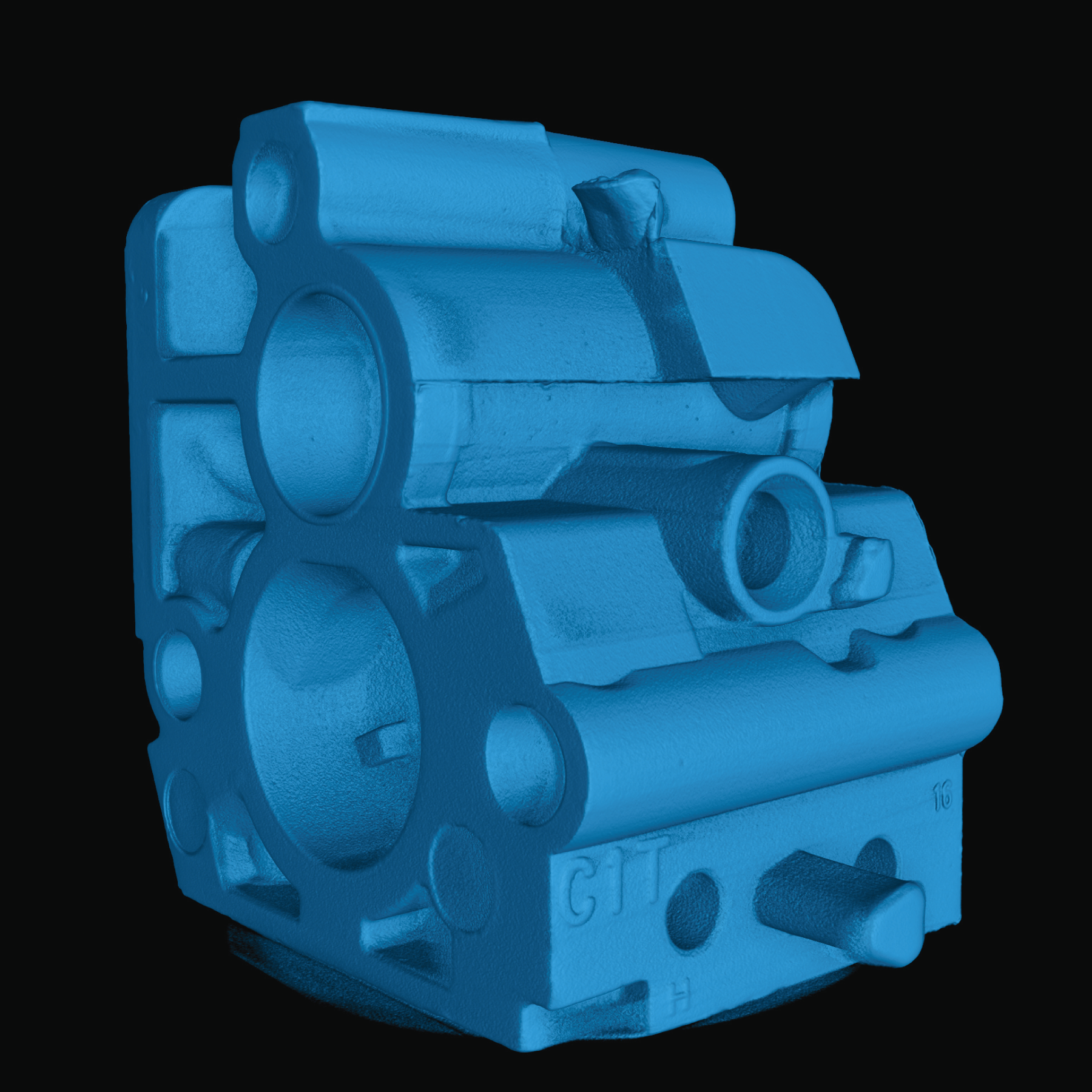
Porosity Inspection in Aluminum Die-Casting
Aluminum die-casting is widely used for automotive parts and electronic enclosures requiring high strength, lightweight properties, and high precision. However, gas entrapment or solidification shrinkage during manufacturing can create microscopic voids, known as porosity. These defects can significantly reduce structural integrity or lead to fluid leakage, making accurate detection and evaluation critical.
Advantages of Industrial CT:Traditional 2D X-ray inspection loses depth information, making it difficult to assess internal defects. Industrial CT scanners capture high-resolution 3D data, allowing for precise visualization of internal porosity.
Data Utilization:Quantitatively evaluating the position, size, and distribution of voids helps engineers optimize casting conditions. This data is essential for establishing accurate defect tolerance standards and improving overall yield.
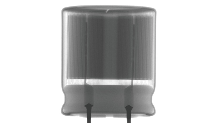
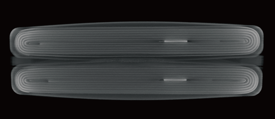
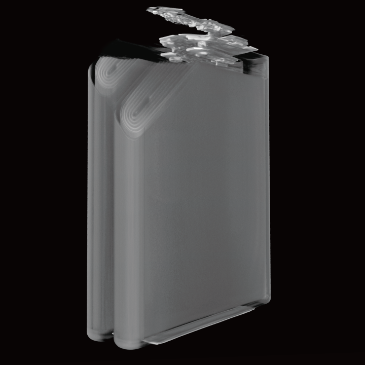
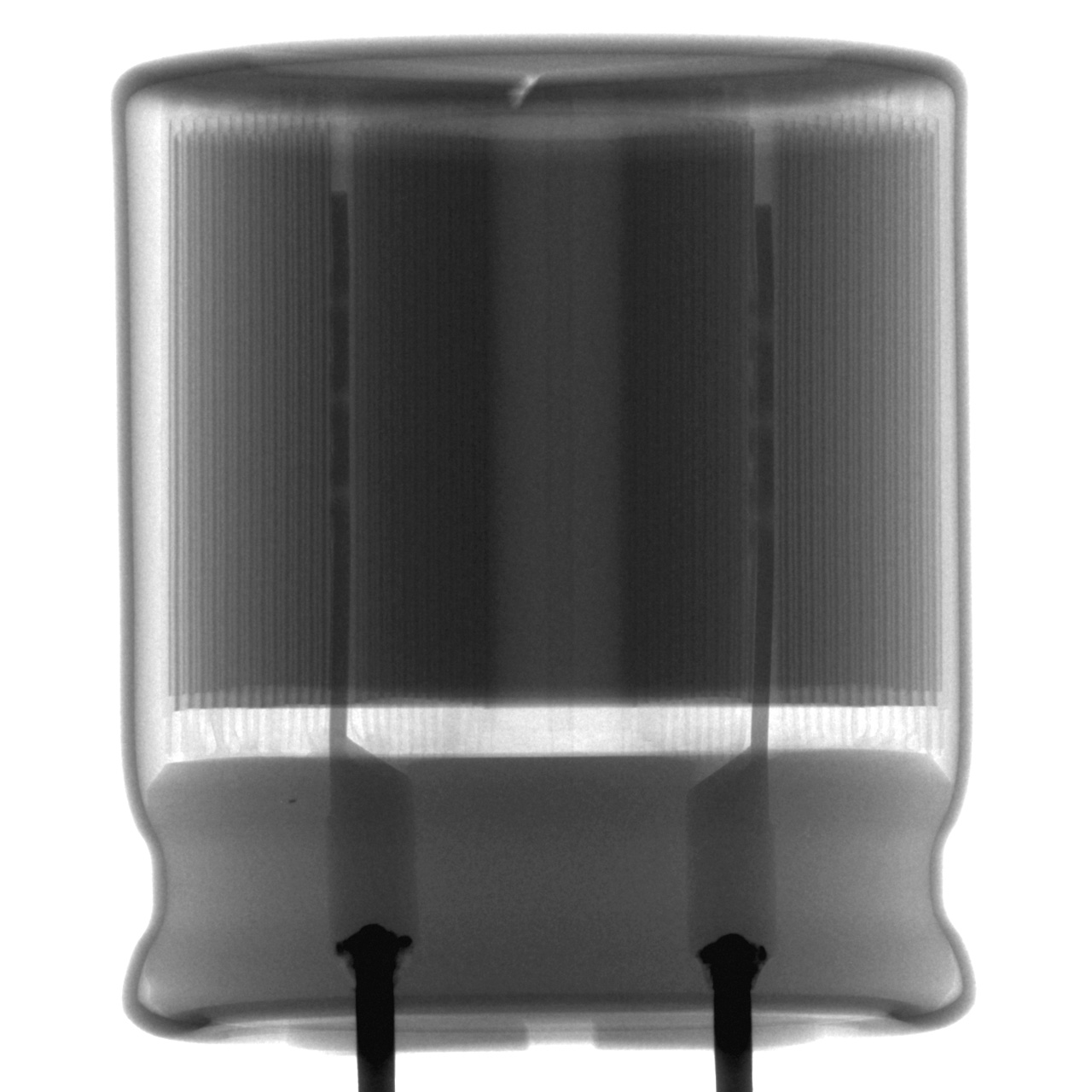
Internal Structure Inspection: Capacitors and Batteries
Electrolytic capacitors rely on a wound structure (jelly roll) of foil electrodes and separators. Precise alignment is essential, as winding shifts can lead to internal shorts or abnormal capacitance.
Advantages of Industrial CT:Industrial CT scanners non-destructively penetrate the outer casing to reconstruct the internal winding or stacked structure in 3D. This allows for the quantitative measurement of the clearance between foils and separators without physical contact.
Ensuring Long-Term Reliability:By analyzing 3D images for winding displacement, stacking distortion, and clearance variations, manufacturers can identify process irregularities early. These inspections ensure the long-term reliability and safety of the finished component.
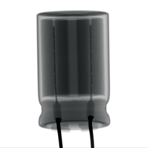
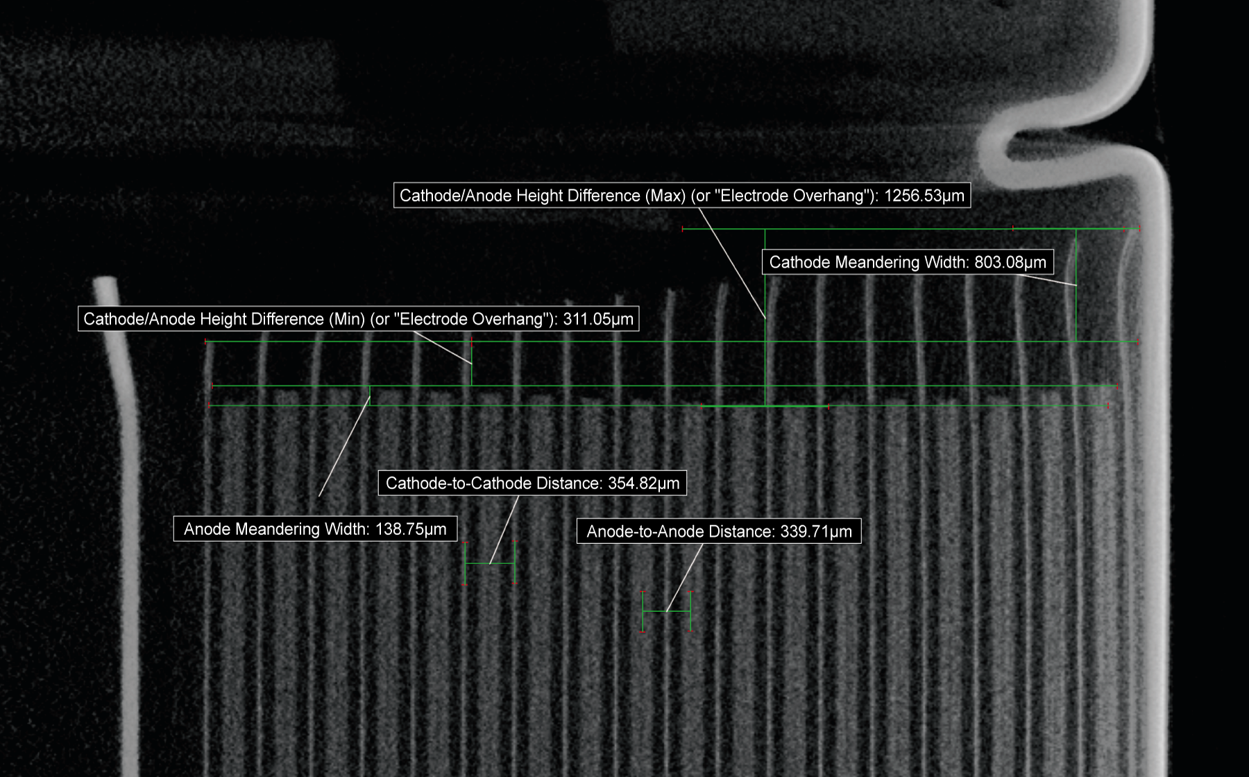
Image Gallery

Datasheets
If you are unable to download a file
Please try the following solution.
- Please press Ctrl+F5 to clear the cache of your web browser and try again.
- Please restart your web browser and log in again to try again.
- Please change your web browser to another browser and try again.
- Restart the computer and try again.
- Please try again on a different computer.
Login Required
-
µRay8700/µRay8760 Datasheet
Date: 2026-06-01 Rev 08
PDF (5,692 KB) -
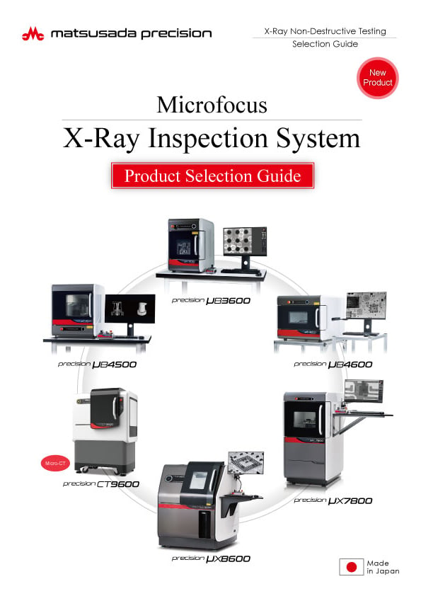
Microfocus X-Ray Inspection System Selection Guide
Date: 2026-05-13 rev 08
PDF (10,805 KB) -
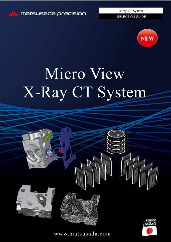
Micro View X-Ray CT System Guide
Date: 2026-05-13 Rev 09
PDF (4,573 KB)
Related Technical Articles
Similar products
-
New
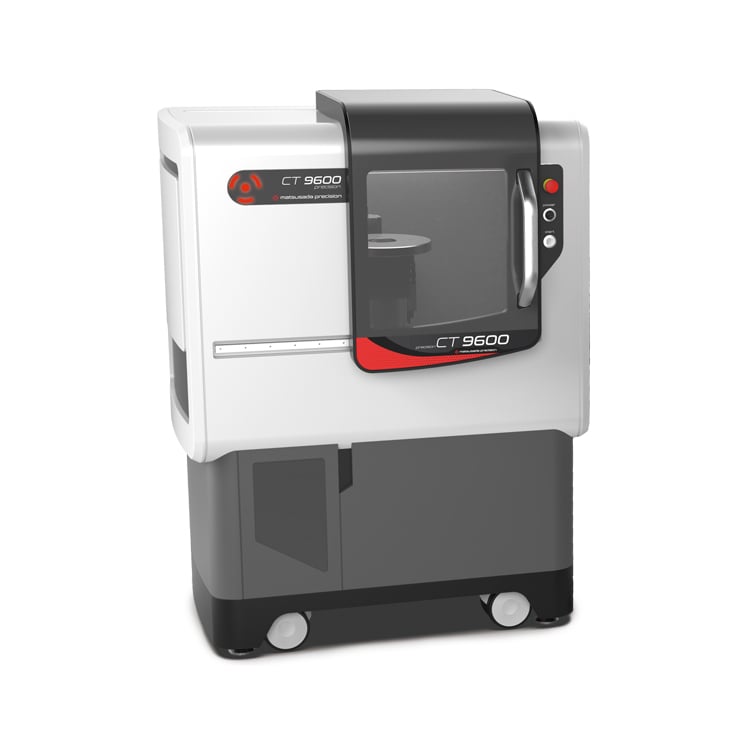
precision CT9600
- X-ray tube voltage
- 130 kV
- X-ray power
- 40 W
Industrial Micro-CT Scanner -
New
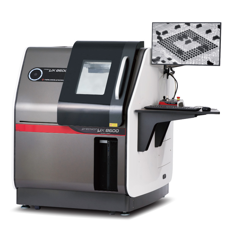
precision μX8600
- X-ray tube voltage
- 130 kV
- X-ray power
- 40 W
Microfocus X-Ray Inspection SystemOptimized for Large Samples and PCBs
-
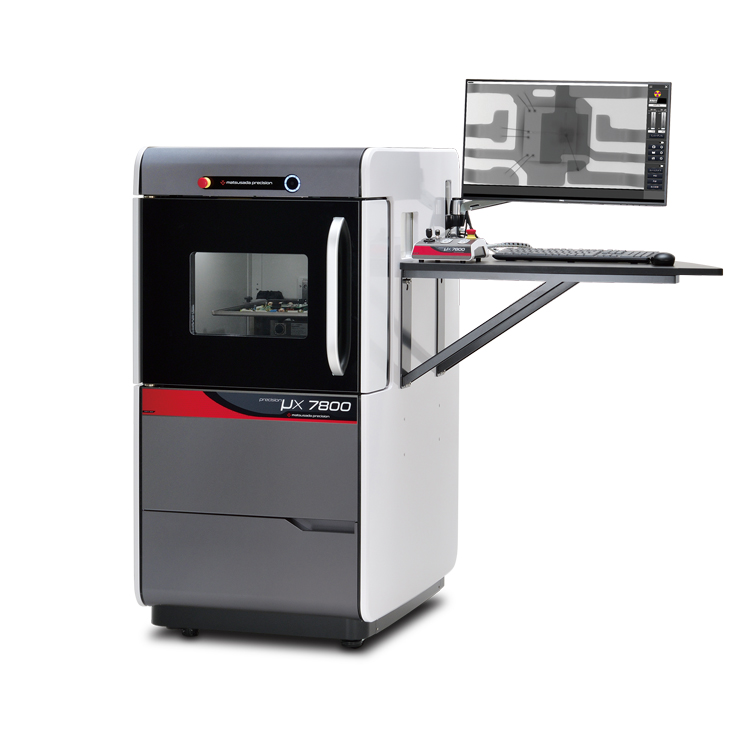
precision μX7800
- X-ray Tube Voltage
- 90 kV
- X-ray Power
- 18 W
X-ray Inspection SystemVersatile General-Purpose Model
-
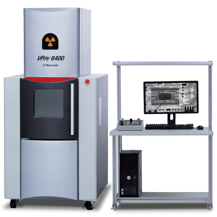
μRay8400
- X-ray tube voltage
- 130 kV
- X-ray power
- 40 W
130 kV micro focus X-raySuitable for observing large footprint material or specimens
Product Lineup:
X-ray Inspection SystemsUser Support
FAQ
- What is Micro‑focus X‑ray?
- What is the tube voltage for X‑ray inspection systems?
- Is a license required for usage of X‑ray inspection system?
- What is the difference between medical and industrial CT scans?
- What is the difference between an X‑ray inspection system and a CT scan system?
- How do we reduce concentric circle noise (ring artifact) in CT images?