Introduction to X-Ray Computed Tomography
X-ray computed tomography (CT) is a powerful non-destructive testing technique that utilizes X-rays to obtain detailed 2D and 3D images. An X-ray CT system radiates the object from multiple directions and analyzes the resulting data to reconstruct a precise three-dimensional model.
By using X-ray CT, an X-ray inspection system provides clear visualization of the internal structure of a component. By capturing X-ray radiation from a single direction, the system can generate two-dimensional images of the object. These X-ray inspection systems are commonly used in medical examinations and airport baggage screening.
There are significant differences between standard X-ray inspection and X-ray CT scanning:
X-ray inspection provides a direct-view image of an object in the direction of the X-ray radiation.
X-ray CT scanning provides cross-sectional images of the specific area radiated by X-rays.
While an X-ray inspection system generates direct 2D vision images, X-ray CT scanning goes further by capturing sliced image data known as tomographs. These slices are then converted through CT image processing on a computer system to create a realistic 3D model.
Principle of X-ray Attenuation
X-ray CT utilizes X-rays as the primary energy source for imaging. Often categorized as "ionizing radiation," X-rays possess extremely short wavelengths and high energy levels, allowing them to interact directly with the atomic structure of an object.
As X-rays penetrate a material, they interact with electrons and other particles orbiting the atoms, resulting in energy loss known as attenuation. The rate of attenuation is significantly higher in dense materials, such as metals, whereas low-density materials like air or paper exhibit minimal attenuation.
In industrial product inspection, this attenuation property allows for the non-destructive evaluation of internal structures that are otherwise invisible to the naked eye. For a given material, attenuation increases with thickness in the direction of the X-ray beam; conversely, thinner sections result in lower attenuation. This fundamental relationship is a key factor in determining the overall quality of computed tomography.
By leveraging these penetration capabilities, CT technology generates high-contrast cross-sectional images for detailed analysis. While standard 2D X-ray images lack depth information (thickness), data acquired through X-ray CT can be visualized from any orientation, providing a comprehensive view of cross-sections at any position within the object.
The following section provides a detailed overview of the X-ray CT system configuration.
X-rays are emitted from the X-ray source to penetrate the object on the stage and are captured by the X-ray detector. The image of the transmitted X-rays acquired by the X-ray detector changes as the object rotates.
An elongated component on the cross-section will be displayed as small in one orientation but large at another angle. This allows CT reconstruction and output of CT image data. In order to reconstruct and analyze the correct picture, software with an appropriate reconstruction algorithm is required.
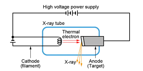
To generate X-rays, a current is first passed through the filament to heat it. The filament generates hot electrons, which are accelerated by high voltage and collide with the target of the anode.
The way computed tomography works is by irradiating the object with X-ray beams from an X-ray source. As the X-ray penetrates the object, it rotates on the rotation stage located between the digital detector panel and the X-ray radiation source. The captured images are reconstructed to develop a 3D model of the part.
Flat Panel Detectors (FPD)
Modern CT systems typically use Flat Panel Detectors (FPD), which are categorized into indirect and direct conversion types.
- Indirect Detectors: Incident X-rays strike a scintillator layer (such as CsI or GOS), converting the X-ray energy into visible light. This light is then detected by a photodiode array and converted into an electrical signal.
- Direct Detectors: These use a photoconductor layer, such as amorphous Selenium (a-Se). When X-rays strike this layer, they are directly converted into electric charges under a high-voltage bias, which are then read out by a Thin Film Transistor (TFT) array.

Direct detectors are based on flat panels of amorphous selenium (a-Se). A high-voltage bias is applied to the selenium film, and a current will flow as X-ray photons hit the film. Then, the current turns on a thin film transistor (TFT), which converts the signal into an image.
Comparison: Medical vs. Industrial CT
There is a big difference between medical and industrial X-ray CT scans.
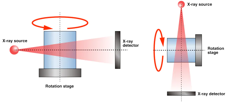
While the underlying physics remains the same, there are significant operational differences between medical and industrial X-ray CT scanning. In medical CT applications, the X-ray source and detector rotate spirally around the patient to capture tomographic slices and generate 360-degree images. This method, known as Helical CT, is necessary because a person cannot be rotated during the scan.
In contrast, an industrial X-ray CT system typically keeps the X-ray source and detector in a fixed position while the object under inspection is rotated. This setup allows for high-precision data collection and the generation of detailed 3D models. Industrial systems are generally classified into two types--fan-beam CT and cone-beam CT--depending on the spread angle of the X-ray beam.
Fan-beam CT scanners utilize X-rays emitted in a thin, fan-shaped spread. This configuration is widely adopted for versatile, high-resolution applications where exceptional image clarity is required.
Cone-beam CT scanners, which project X-rays in a conical shape, are highly popular in industrial sectors. A key advantage of the cone-beam system is its ability to reconstruct full 3D volume data from a single rotation, significantly improving inspection efficiency.
To meet diverse inspection requirements, cone-beam CT systems support various geometric scanning methods, including Normal, Full, Half, Offset, Continuous, and Intermittent scans.
Industrial CT Applications
Industrial CT scanning technology is increasingly adopted across various sectors, primarily for quality control (QC) and research and development (R&D). In QC applications, for example, X-ray inspection systems provide critical failure analysis for aluminum castings that do not meet structural requirements. The technology effectively detects internal defects, such as shrinkage cavities, ensuring product integrity.
Industrial CT scanning enables comprehensive non-destructive inspection (NDI) to identify:
- Internal structures and material flaws
- Foreign contaminants
- Shrinkage cavities caused by defective filling
- Root causes in failure investigations
- Dimensional data for reverse engineering
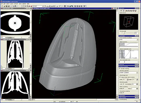
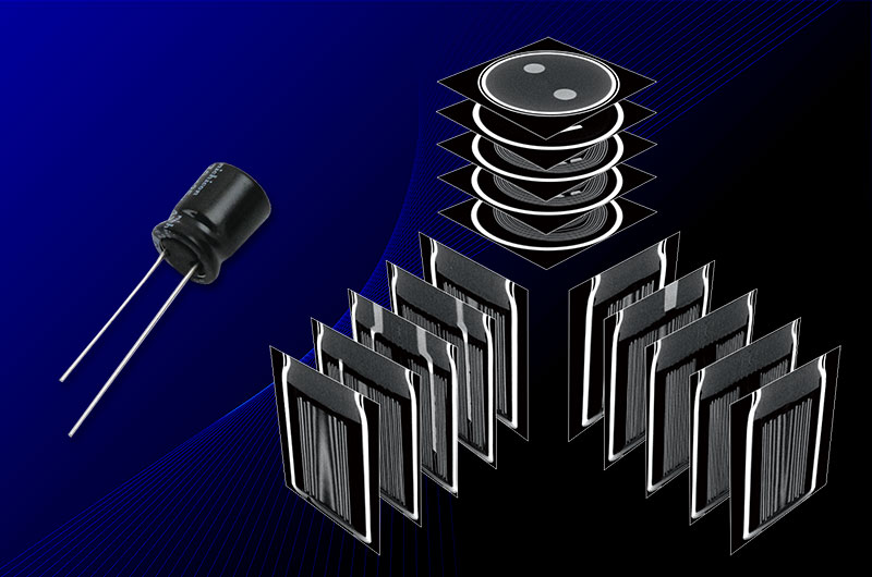
Example of X-ray Computed Tomography Analysis
In R&D, industrial CT solutions are essential for the characterization of high-performance materials and complex structures, including:
- Microdevices and microstructures
- Glass Fiber Reinforced Plastics (GFRP) and composite materials
- Advanced polymers and porous metals
Beyond industrial sectors, X-ray CT is a valuable tool in fields such as archaeology. The non-destructive nature of industrial X-ray CT allows researchers to examine the internal structures of ancient artifacts without compromising their integrity.
Our Micro-CT and 3D microfocus X-ray systems are engineered to provide the high-resolution imaging required for these detailed analyses. These products deliver precise 3D visualization comparable to standard industrial and medical CT systems, offering ultra-high resolution to visualize even the fine pores within porous ceramics.
For detailed specifications or to discuss your specific application, please contact us. Custom configurations are available upon request.
Related Technical Articles
- Principles of Radiography
- How to Acquire High-Quality Computed Tomography (CT) Images - X-ray NDT series (1)
- A Guide to X-ray CT Images: Formats, Viewing, and Applications - X-ray NDT series (2)
- X-ray Image Processing and Automated Inspection - X-ray NDT series (3)
- FAQ: What is the tube voltage or acceleration voltage necessary for X-ray inspection systems?
Recommended products
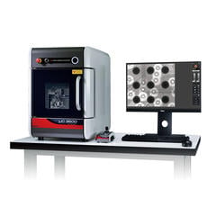
Matsusada Precision's X-ray CT Scanners can capture high-definition and high-resolution images with its unique microfocus X-ray technology.