


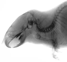
High-resolution X-ray imaging is essential for observing fine skeletal structures and minute density changes in biological specimens.
The μRay8700-LF6 features a 6-megapixel high-resolution Flat Panel Detector (FPD) with a wide field of view, capable of capturing up to 81.6 x 104.1 mm in a single shot. This allows for efficient imaging of entire laboratory mice without the need for image stitching.
Furthermore, the optional CT capability enables non-destructive cross-sectional analysis, allowing researchers to visualize internal bone structures in three dimensions.
X-ray system parameters
| Focal spot | Microfocus |
|---|---|
| X-ray tube voltage | 30 to 90 kV |
| Magnification | 1 to 10x |
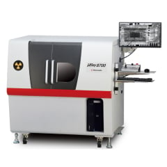
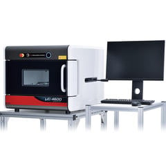
Recommended products

Related Technical Articles
- Selecting an X-ray Inspection System
- Principles of Radiography
- Safe Operation of X-ray Inspection Systems
- What is Microfocus X-ray Technology? (Basic Knowledge)
- Basics and Principles of Computed Tomography (CT)
- What are X-rays? (Basic Knowledge)
- How to Acquire High-Quality Computed Tomography (CT) Images - X-ray NDT series (1)
- A Guide to X-ray CT Images: Formats, Viewing, and Applications - X-ray NDT series (2)
- X-ray Image Processing and Automated Inspection - X-ray NDT series (3)
- Types of X-ray Tubes and High-voltage Power Supplies
- What is the difference between Radioactivity, Radiation, and Radioactive Materials?
- Understanding Radiation: Effects on the Body and X-ray Safety