


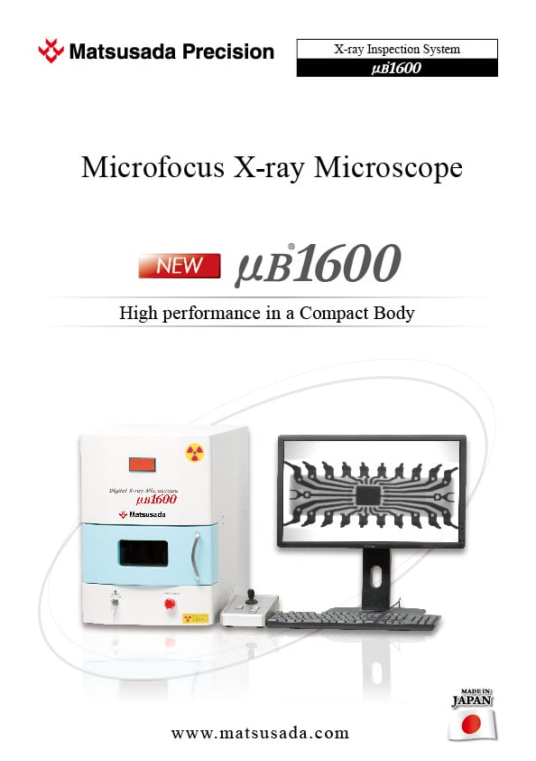
Microfocus X-ray
Microscope
- X-ray Voltage:
20 kV to 60 kV - Microfocus x-ray tube
High Performance in a Compact Body
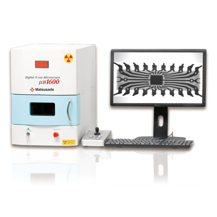
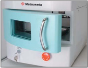
Leveraging our expertise in high-voltage power supply technology, Matsusada Precision presents the µB1600, a compact benchtop microfocus X-ray inspection system. Featuring an internal microfocus X-ray source and a high-resolution camera, the µB1600 delivers precise transmission imaging and measurement capabilities in a space-saving design suitable for any installation environment.
Features and Benefits
Hardware
- Microfocus X-ray tube
- Digital imaging camera
- 4.7" x 4.7" stage area
- X-Y shifting stage and Rotating stage
- Joystick controller
- Low-leakage X-ray dose (less than 1 µSv/hr)
- Safety functions: Interlock, emergency stop, and more
Software
- 14-bit density resolution
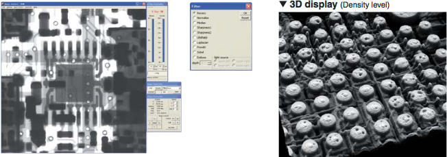
- Density processing: 3D visualization, false color, pseudo-3D
- Intensity processing: Brightness, contrast, non-linear contrast stretch (gamma), filtering, inversion, binarization, histogram
- Measurement functions: Distance, angle, and area
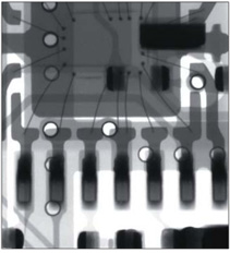
Applications
- Composites, ceramics and plastics
- Electronic and electrical components
- Populated and unpopulated Pcb's
1. Advanced Technology Integration
High-resolution imaging driven by state-of-the-art X-ray and FPD technology
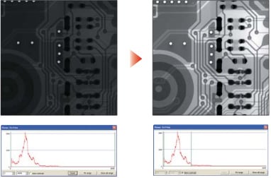
Matsusada Precision’s proprietary technology detects subtle density contrasts often missed by conventional devices. The built-in Flat Panel Detector (FPD) ensures distortion-free imaging for accurate analysis.
2. Intuitive Operation
Automatic optimization of imaging conditions
Operators simply place the specimen and activate the X-ray. The µB1600 automatically adjusts contrast and exposure settings to capture the specimen under optimal conditions.
3. Versatile Functionality
Standard inclusion of advanced image processing and measurement software
The µB1600 is equipped with extensive functions suitable for both analysis and routine inspection. The user-friendly interface ensures smooth operation, even for inexperienced operators.
Leading Edge Technology in compact body
World's FirstMicrofocus X-ray
The µB1600 has an internal microfocus X-ray source creating ultra-high image quality in spite of its compact size. The integration of 60kV and 150µA X-ray tube and Matsusada Precision's high-voltage technique has succeeded in developing a small-sized and high-performance X-ray generator.
Digital X-ray camera
A digital X-ray camera with a flat panel is used in the imaging part. The high-resolution camera with one million pixels of 1024 ×1024 clearly detects an object aimed at. Images with no distortion in every detail can be obtained.
14-bit digital data
High-definition data with 1.5 million pixels are imported by 14-bit. The images can be processed and analyzed flexibly afterward.
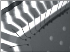
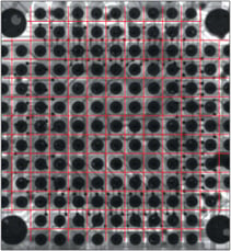
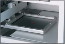
High-precision stage
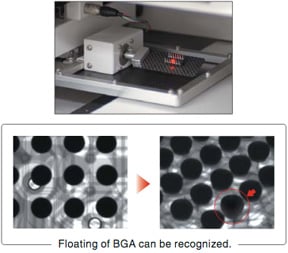
The series has the rotatable stage along with the three axes of X, Y axes and variable enlargement ratio axis, enabling transmission from any angle. Moreover, it meets your needs for more detailed imaging for smaller parts. It can also detect floating BGAs.
Model
| Model | X-ray Source | Imaging Section | Stage | ||||||||||
|---|---|---|---|---|---|---|---|---|---|---|---|---|---|
| Anode voltage | Anode current | Focal size | Size of visual field | Number of valid pixel | Density resolution | Monitor | Lower and upper imaging part Stroke | Sample platform | XY Stroke | Rotation Stroke | Shifting operation | ||
| Size | Load capacity | ||||||||||||
| μB1600 | 20 kV to 60 kV | 150 µA MAX | 5 µm | 2.52 inches (64 mm) x 2.24 inches (57 mm) | 1.5 M pixels | 14-bit | 24 inches | 7.87 inches (200 mm) | X 4.72 x Y 4.72 inches (X 120 x Y 120 mm) |
1kg | X 4.72 x Y 4.72 inches (X 120 x Y 120 mm) |
360° (manual) | Direction indication by mouse, Joystick can be used together. |
| Geometric image magnification | 11 to 19.8 times |
|---|---|
| Monitor magnification | 60 to 100 times |
| Field of view | 0.16 x 0.20 inches (3.2 x 3.6 mm) to 0.24 x 0.28 inches (5.0 x 6.0 mm) |
| Input voltage | 100 Vac 50/60 Hz * |
| Dose of X-ray leakage | less than 1 µSv/hr |
* The other voltage is selectable. Please ask the sales department.
Functions
Ultimate Operability
Automatic conditioning function
The system tracks usage history and automatically selects the appropriate conditioning process based on the elapsed time since the last operation. This ensures optimal performance and significantly extends the life of the X-ray tube.
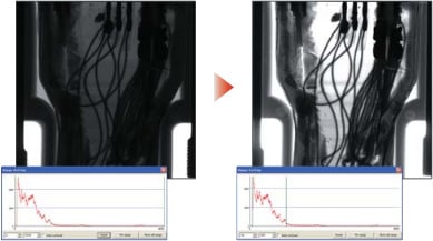
Auto contrast adjustment function
Images shot by 14-bit are displayed in optimal gradation sequence. Variations by photographers (e.g., dark images, whitish images, etc) can be eliminated to always display optimally. Even a first-timer does not miss the points that he or she wants to observe. It is also possible to display any given tonal range.
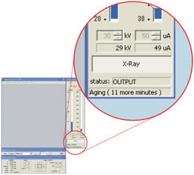
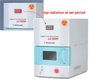
Automatic Shutoff Function
To prevent accidental continuous operation, the automatic shutoff function turns off the X-ray output after a pre-set time. This feature enhances safety and energy efficiency if the operator leaves the device unattended.
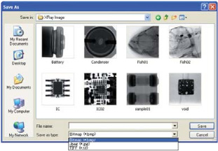
Data storage (bmp, jpg, tif)
Images both before and after analyses can be easily saved. Extensions including bmp, jpg, and tif can be selected depending on the extendability. Once saved data can be read out and analyzed repeatedly by the analysis software.
Smooth & Easy Operation even for first-timers
Image analysis
The brand new image analysis software dedicated for our x-ray inspection system can controls the stage and X-ray with easily operations and is equipped with various image processing and measurement functions.
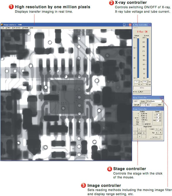
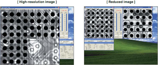
Reduced display function
The moving image window displays not only high-resolution images with one million pixels but also reduced images allowing specimens to be inspected compared with the images on the static image window.
Filter function
The µB1600 supports ten or more types of filtering and image processing, allowing detailed image analyses. Images can be analyzed in real-time by performing appropriate filtering for displaying moving images.
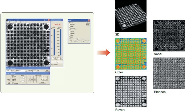
Image analyze window function variety
Display range window
The contrast can be enhanced manually by setting any given densities based on the histogram of shot images.
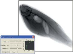
Brightness adjustment
Brightness values of shot images can be adjusted freely by adjusting gain, contrast and gamma values individually.
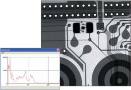
Histogram display
The µB1600 displays a histogram to indicate the distribution of brightness of shot images.
Binarizing processing
A shot image can be displayed in a binary manner by contrast by setting a threshold and binarizing the image.
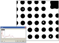
Area calculating
The µB1600 can measure the areas, number of holes, area of holes and area ratio of an object in a shot image.
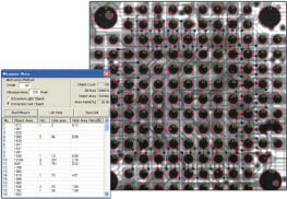
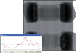
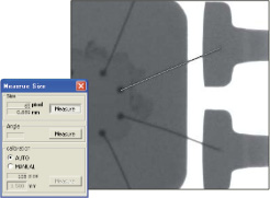
Size measurement
Sizes in a captured image can be measured by drawing measurement lines on the shot image.
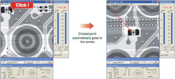
Shift by click
Click on an image, and quickly move to the location you want to observe.
In addition to normal stage operation, the clicked section becomes centered.
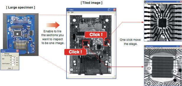
Stitching Function (Interlink)
The Interlink function allows for the capture of large samples (up to 4.72" x 4.72") that cannot be viewed in a single shot. By stitching multiple images together, the system creates a complete overview of the sample. Users can then navigate to specific points of interest simply by clicking on the stitched image.
Teaching function
The teaching function moves the stage automatically to the pre-registered points. This allows the system to "learn" problem areas and capture them on an X-ray image automatically. Setting X-ray output, enlargement factor and stop time, etc., for each point supports various test objectives. The teaching function moves the stage automatically to the pre-registered points. This allows the system to "learn"
For various applications
Electrical components
BGA
Capacitor
Liquid crystal device
Electrical components
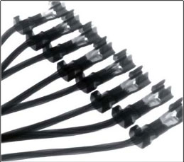
Connector
Metal parts
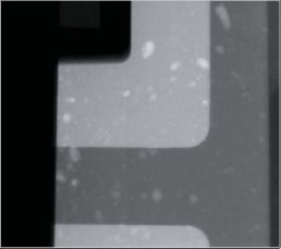
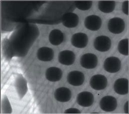
Holes in Aluminum sample
Biological specimens
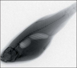
X-ray image of a fish
Various standard functions
Easy operations by joystick
The joystick allows easy movements of the stage along the X and Y axis, 360-degree rotation of the stage, and changes in image magnification.
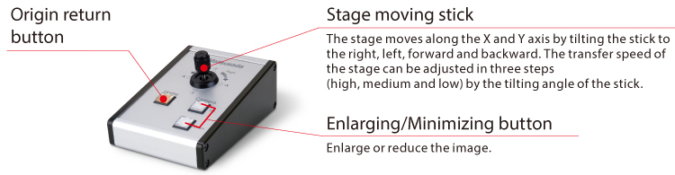
Display the position of observing
The laser pointer indicates the center of the displayed image. It is possible to position by observing the sample.
Safety functions
- Low-leakage X-ray Dose
- Designed with comprehensive shielding, the µB1600 maintains surface radiation levels below 1 µSv/hr, simplifying safety management and installation requirements.
(Note: In some regions, special licensing may not be required.)
- Emergency Stop Function
- Pushing this button stops the x-ray and the stage from operating in an emergency situation.
- Interlock Function
- The X-ray will shut off the moment the door is opened.
Specifications
Accessories
- XDSK100
Aluminum table for option
- STG002
Height adjustment stage
-
Size: 120 mm × 120 mm
The stage helps you to raise its height. Therefore, the sample can be closer to the X-ray camera for shooting, so you can take larger field images.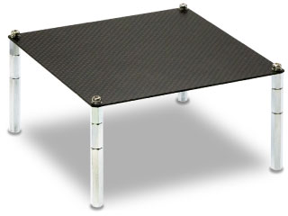
Applications
Fields and Applications of X-ray Radiography Non-Destructive Testing
- Pharmaceuticals and Cosmetics
- Tablets, Capsules, Catheters, Medical pouches, Foreign objects in the face mask, Cosmetics
- Automobiles and Electric Vehicles
- Automotive components, Wire harness, Batteries, Molded plastic parts, Motor, Insert molded parts, Aluminum die casting, Sensor, Mating parts connection
- Electronics
- Ball grid array (BGA), Integrated circuit (IC) chip, Connectors, Capacitors, LED, Relays, Printed circuit board (PCB), AC adapter, Light bulbs filament, Electrical cable disconnects, Discharge traces of dielectric breakdown
- Advanced Materials
- Carbon-Fiber-Reinforced Polymers (CFRP), Insulation material, Carbon, Dissimilar material joint, Contamination in beryllium
- Precision Components
- Probe, Bearing, Mating of plastic parts, Spring position
- Biological Samples (Creatures and Plants)
- Laboratory rats, Seeds, Flowers, Shell, Bone, Insects, Rice
- Others
- Microscopic foreign matter contamination, Voids in welding, Plastic bottle, Sealing of cans
Download
If you are unable to download a file
Please try the following solution.
- Please press Ctrl+F5 to clear the cache of your web browser and try again.
- Please restart your web browser and log in again to try again.
- Please change your web browser to another browser and try again.
- Restart the computer and try again.
- Please try again on a different computer.
-
μB1600 Datasheet
Date: 2026-04-22 Rev 09
PDF (4,635 KB)
Login Required
-
μB1600 Datasheet
Date: 2026-04-22 Rev 09
PDF (4,635 KB)
On this website, we provide only the latest versions of information and instruction manuals for our products. Therefore, the newest versions of manuals on the website may differ from those that came with products you purchased in the past.