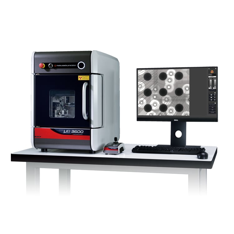
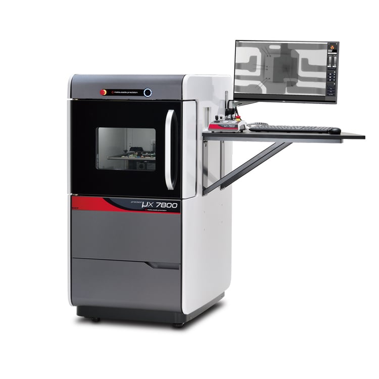
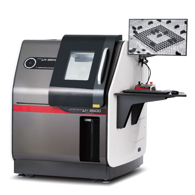
The term "X-ray microscope or X-Ray Microscopy (XRM)" was originally used to describe a method for observing biological samples that contain moisture at synchrotron radiation facilities such as Spring-8. This is achieved by using synchrotron radiation with a wavelength shorter than that of visible light to obtain observation images with high spatial resolution. X-ray microscopes at synchrotron radiation facilities are large devices with long optical paths, using monochromators, multilayer focusing mirrors, rotating elliptical mirrors, and zone plates for focusing and imaging X-rays.
Recently, the following devices, which do not use synchrotron radiation, are also called "X-ray microscopes". This is a device that uses X-rays to magnify (photograph) internal structures. Since X-rays cannot be seen directly by the eye, X-rays irradiated to a sample are magnified and projected onto a fluorescent plate, etc., converted into visible light, and photographed, or photographed by a camera for X that converts X-rays directly into signals, and made into images.For the following instruments, magnification and resolution are comparable to those of optical microscopes: magnification: up to 1000x, resolution: limit to 0.2μm.


