Positron emission tomography (PET) is a nuclear medicine imaging technique that visualizes metabolic processes within the body. Unlike CT scans, which primarily provide detailed anatomical images, PET detects pairs of gamma rays emitted indirectly by a positron-emitting radiopharmaceutical (tracer), such as Fluorodeoxyglucose (FDG). This tracer accumulates in cells with high metabolic activity, allowing for the effective detection of tumors and metastasis. Consequently, PET is widely used in oncology for cancer screening and treatment monitoring, as well as in neurology for diagnosing conditions like epilepsy and Alzheimer's disease.
Modern systems often combine PET with CT (PET/CT) to capture both functional and anatomical data in a single session, providing comprehensive diagnostic information while managing radiation exposure.
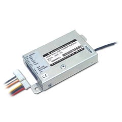
Detectors and High-Voltage Power Supplies
PET scanners utilize a ring of detectors containing scintillators that convert gamma rays into light. This light is captured by photodetectors such as Photomultiplier Tubes (PMTs), Avalanche Photodiodes (APDs), or Silicon Photomultipliers (SiPMs), which convert it into electrical signals for image reconstruction. These detectors require highly stable, low-noise high-voltage power supplies (typically ranging from 100 V to 5 kV) to ensure precise measurement and high-quality imaging.
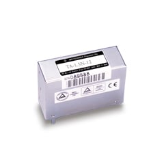
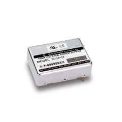
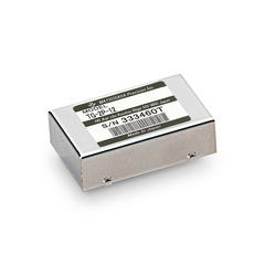
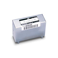
Matsusada Precision offers a wide range of compact, high-reliability high-voltage power supplies optimized for PET detectors.
How a PET-CT system works
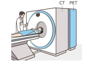
The system integrates PET and CT technologies, operating simultaneously to generate fused images that provide both functional and anatomical information.
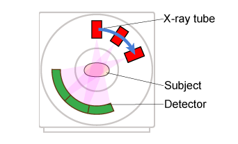
Data is collected by rotating the detector opposite the X-ray tube.
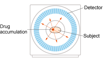
Detectors arranged in a ring capture radiation emitted from the radiopharmaceutical inside the patient's body.
- Related Terms:
-
- Image Intensifier (I.I.)
- Photomultiplier Tube (PMT)
- Avalanche Photodiode (APD)
- Microchannel Plate (MCP)
- Scintillator
- Radiation
- CT
- Positron Emission Tomography (PET)-Scanner
Recommended products
We offer a variety of high-voltage power supplies for detectors of Positron Emission Tomography (PET).