


Flow cytometry is an essential technique for cell analysis. This article describes the fundamental principles of flow cytometry, the instrumentation involved, the mechanism of cell sorting, and its common applications.

What is flow cytometry?
Flow cytometry is a powerful analytical technique used to measure physical and chemical characteristics of a population of cells or particles. The process involves preparing the sample as a single cell suspension in a fluid and injecting it into a flow cytometer. As the cells flow single-file through a laser beam, the instrument analyzes individual cells at high speed.
This technique measures various parameters simultaneously, including cell size, internal complexity, and fluorescence intensity. It provides detailed, quantitative data on heterogeneous cell populations, making it essential for research and clinical diagnostics.
Understanding the Flow Cytometer
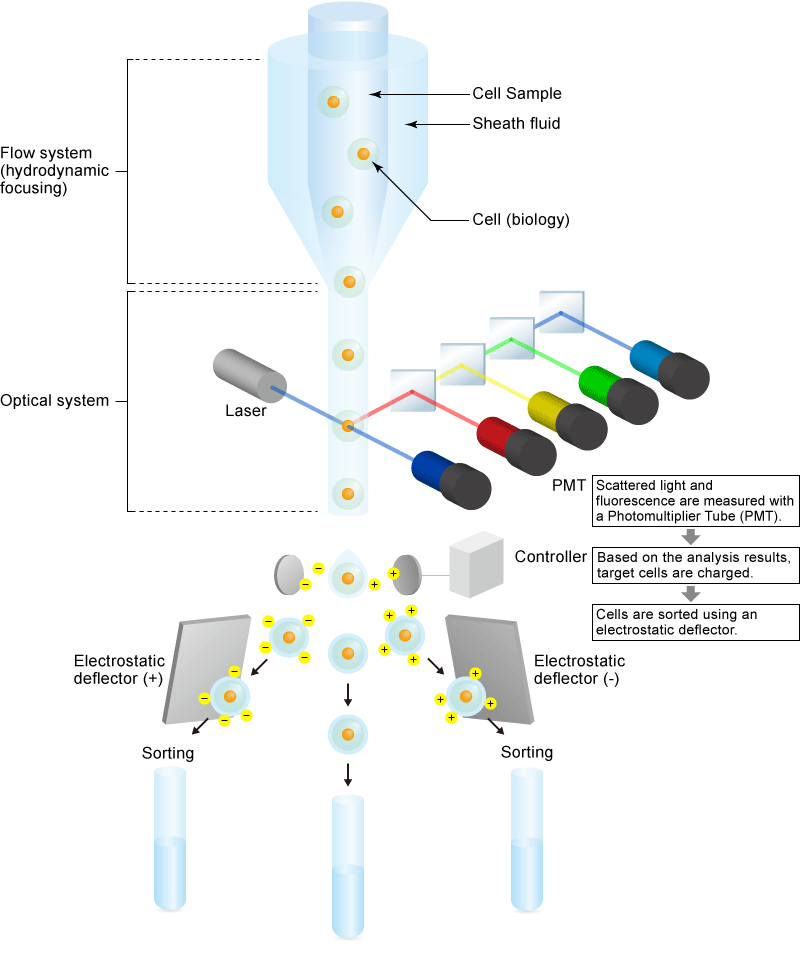
A flow cytometer integrates three main systems: fluidics, optics, and electronics.
- Fluidics System: Transports cells in a stream to the laser beam for interrogation. Using a sheath fluid, the system applies hydrodynamic focusing to align cells in a single file.
- Optics System: Consists of lasers to illuminate the particles and optical filters to direct the emitted light to detectors. Photomultiplier Tubes (PMTs) are typically used to detect light scattered by the cells and fluorescence with high sensitivity.
- Electronics System: Converts the analog light signals detected by the PMTs into digital data for computer analysis.
Cell Sorting Mechanism
Some flow cytometers, known as cell sorters, can physically isolate specific cells of interest from a mixed population. Based on the analysis of each cell, the instrument applies a precise electrical charge to the droplet containing the target cell. As the droplet passes through high-voltage deflection plates, the electrostatic field deflects it into a collection tube. This allows for the non-destructive recovery of cells for further culture or analysis.
Experimental Design Workflow
Proper planning is critical for successful flow cytometry experiments.
- Fluorochrome Assignment: Carefully assign fluorescent dyes or fluorescent proteins to antibodies or cell markers to minimize spectral overlap (spillover). Consider the excitation/emission spectra of the dyes and the configuration of the instrument's lasers and filters.
- Sample Preparation and Staining: Select the appropriate staining protocol (surface, intracellular, or indirect staining) based on the target application.
- Filtration: Filter samples immediately before analysis to remove cell clumps. Aggregates can clog the fluidics system and compromise data integrity.
Data Analysis and Applications
Flow cytometry is widely used in immunology, molecular biology, and cancer research. It enables the detection of cell surface markers, intracellular cytokines, and gene expression (e.g., using GFP).
In clinical settings, it is indispensable for analyzing blood cells to diagnose hematological disorders such as leukemia and for monitoring immune status (e.g., CD4 counts in HIV). Compared to microscopy, flow cytometry offers high-throughput capability--analyzing thousands of cells per second--and provides highly quantitative, multi-parametric data.
- Related Terms:
Reference (Japanese site)
- 高分子学会 - フローサイトメトリー(Flow Cytometry)
(https://www.spsj.or.jp/equipment/news/news_detail_42.html) - Thermo Fisher - フローサイトメーターの仕組み
(https://www.thermofisher.com/jp/ja/home/life-science/cell-analysis/cell-analysis-learning-center/molecular-probes-school-of-fluorescence/flow-cytometry-basics/flow-cytometry-fundamentals/how-flow-cytometer-works.html) - Cell Signaling - フローサイトメトリーの概要
(https://www.cellsignal.jp/applications/flow-cytometry/flow-cytometry-overview)